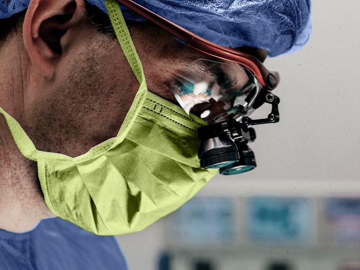
Brain surgery is any operation or procedure that treats problems involving the brain, skull, blood vessels around the brain, protective membranes, or cerebrospinal fluid pathways. That sounds like something from a high-stakes medical drama, complete with dramatic lighting and someone saying, “We need ten more minutes.” In real life, brain surgery is usually far more planned, precise, and team-based than television makes it look.
Neurosurgeons perform brain surgery to remove tumors, repair abnormal blood vessels, drain excess fluid, relieve pressure, treat epilepsy, control movement disorder symptoms, collect a biopsy, or repair damage after bleeding, stroke, infection, or injury. Some procedures require opening part of the skull. Others use small openings, endoscopes, image guidance, lasers, catheters, or implanted devices. The goal is not simply to “operate on the brain,” but to solve a specific problem while protecting speech, movement, memory, vision, balance, and personalitythe things that make a person feel like themselves.
This guide explains the major types of brain surgery, what patients can expect before and after a procedure, possible risks, and what recovery often looks like at home. It is educational, not a substitute for medical advice, because your brain deserves more than a random internet pep talk.
What is brain surgery?
Brain surgery, also called neurosurgery when performed by a neurosurgeon, includes procedures that treat diseases or injuries affecting the brain and nearby structures. The exact technique depends on the diagnosis, the location of the problem, the person’s overall health, and whether the situation is planned or urgent.
For example, a person with a slow-growing benign brain tumor may have a carefully scheduled operation after several scans and consultations. Someone with life-threatening bleeding or severe brain swelling may need emergency surgery to relieve pressure. A person with Parkinson’s disease may have deep brain stimulation to help reduce movement symptoms, while a person with hydrocephalus may receive a shunt to drain excess cerebrospinal fluid.
Why brain surgery may be needed
Brain surgery may be recommended when medication, monitoring, radiation, or other treatments are not enough. Common reasons include:
- Brain tumors: Surgery may remove all or part of a tumor, reduce pressure, confirm a diagnosis, or make other treatments more effective.
- Brain aneurysms: Some aneurysms may require clipping or endovascular treatment to lower the risk of bleeding.
- Bleeding or blood clots: Surgery can remove blood, relieve pressure, or repair the source of bleeding.
- Epilepsy: Surgery may remove or disconnect the area causing seizures when medications do not control them.
- Hydrocephalus: A shunt or endoscopic procedure may help drain excess fluid.
- Movement disorders: Deep brain stimulation may help selected people with Parkinson’s disease, essential tremor, dystonia, or other conditions.
- Traumatic brain injury: Surgery may be needed to reduce swelling, remove blood, or repair skull damage.
- Infection or abscess: Some infections require drainage or removal of infected tissue.
Common types of brain surgery
1. Craniotomy
A craniotomy is one of the best-known types of brain surgery. During this procedure, the surgeon temporarily removes a section of the skull, called a bone flap, to access the brain. After the operation, the bone flap is usually replaced and secured.
Craniotomy may be used for brain tumors, aneurysms, blood clots, epilepsy surgery, vascular malformations, or pressure relief. The location of the opening depends on the condition. For example, a surgeon may use a small eyebrow incision to reach certain tumors or aneurysms near the front of the brain, or an opening behind the ear for specific skull-base problems. Modern craniotomy often uses navigation systems that work somewhat like GPS, except the destination is not a coffee shopit is a millimeter-level target inside the skull.
2. Craniectomy and cranioplasty
A craniectomy is similar to a craniotomy, but the removed bone is not replaced right away. This may be done when the brain is swollen and needs room to expand safely. The bone may be stored and replaced later, or a custom implant may be used. The later repair procedure is called cranioplasty.
Craniectomy is often associated with urgent situations such as traumatic brain injury, stroke-related swelling, or severe bleeding. It is not a casual operation; it is usually done to protect the brain from dangerous pressure.
3. Brain biopsy
A brain biopsy removes a small sample of tissue so specialists can study it under a microscope. Doctors may recommend a biopsy when imaging shows an abnormal area but the diagnosis is unclear. Some biopsies use a stereotactic frame or advanced image guidance to reach the target through a small opening.
The information from a biopsy can help determine whether a lesion is cancer, inflammation, infection, scar tissue, or another condition. In other words, it helps the medical team stop guessing and start planning.
4. Awake brain surgery
Awake brain surgery, often called awake craniotomy or awake brain mapping, may be used when a tumor or seizure focus is close to areas controlling speech, movement, or other critical functions. The patient is not simply “left awake” like someone forgot the anesthesia. Instead, the anesthesia team carefully manages comfort and sedation, and the surgical team may ask the patient to speak, move, count, name pictures, or answer questions during mapping.
This technique helps surgeons identify important brain areas and avoid them when removing abnormal tissue. It can sound intimidating, but for selected patients, it may improve safety and preserve function.
5. Endoscopic and minimally invasive brain surgery
Some brain procedures use an endoscope, a thin tube with a camera and small instruments. Endoscopic surgery may require a smaller opening than traditional surgery and can be used for certain tumors, cysts, fluid problems, or skull-base conditions.
Minimally invasive techniques do not mean “minor.” They still involve the brain and require expert care. However, when appropriate, they may reduce tissue disruption, shorten hospital stays, and speed certain parts of recovery.
6. Laser interstitial thermal therapy
Laser interstitial thermal therapy, or LITT, uses image guidance to place a laser probe into abnormal tissue, often through a small opening. Heat from the laser can destroy targeted tissue, such as certain tumors or seizure-causing areas. It may be an option for patients who are not good candidates for open surgery or whose lesion is deep or difficult to reach.
LITT is not right for every brain condition, but it shows how neurosurgery has moved far beyond the old idea that every brain operation requires a large opening.
7. Deep brain stimulation
Deep brain stimulation, or DBS, involves implanting thin electrodes in specific brain areas. These electrodes connect to a pulse generator, usually placed under the skin in the chest. The device sends controlled electrical signals that can help regulate abnormal brain activity.
DBS is used for selected people with Parkinson’s disease, essential tremor, dystonia, and certain other neurological conditions. It does not cure the underlying disease, but it may reduce symptoms and improve quality of life. After surgery, the device must be programmed and adjusted, so DBS is more like getting a smart thermostat for brain circuits than flipping a simple on-off switch.
8. Shunt surgery for hydrocephalus
Hydrocephalus occurs when cerebrospinal fluid builds up in the brain’s ventricles. A shunt is a flexible tube system that drains extra fluid from the brain to another part of the body, often the abdomen, where it can be absorbed. A valve helps regulate the flow.
Shunt surgery may improve walking, thinking, bladder control, headaches, or pressure-related symptoms, depending on the cause of hydrocephalus. Some people improve quickly, while others take weeks or months. Shunts also require long-term monitoring because they can malfunction, become blocked, or become infected.
9. Aneurysm surgery and neurovascular procedures
A brain aneurysm is a weak bulge in a blood vessel wall. If treatment is needed, options may include surgical clipping through a craniotomy or endovascular procedures performed through blood vessels. Clipping places a tiny clip at the base of the aneurysm to keep blood from entering it. Endovascular treatment may use coils, stents, or flow-diverting devices.
Not every aneurysm needs immediate treatment. Doctors weigh size, location, symptoms, rupture risk, age, health, and patient preferences before recommending a plan.
What to expect before brain surgery
Before brain surgery, patients usually meet with a neurosurgeon, anesthesia team, and sometimes neurologists, oncologists, rehabilitation specialists, or other clinicians. The pre-surgery plan may include MRI, CT, angiography, blood tests, heart testing, medication review, and a discussion of benefits and risks.
Patients are often asked to stop certain medications, especially blood thinners, before surgery if their doctor says it is safe. They may need to avoid eating or drinking for a specific period before anesthesia. The team will explain whether hair needs to be clipped, where the incision may be, how long the hospital stay may be, and what kind of recovery help may be needed at home.
One of the most helpful things patients can do is prepare questions. Good questions include: What is the goal of surgery? What are the alternatives? What functions are near the surgical area? What symptoms should improve? What symptoms may be temporary or permanent? When can I drive, work, exercise, fly, or return to school? A prepared patient is not annoying; a prepared patient is a gift to the care team.
What happens during brain surgery?
The details vary widely. Some people receive general anesthesia and are fully asleep. Others have awake mapping with periods of sedation and interaction. Some procedures use a large operating room team, including neurosurgeons, anesthesiologists, nurses, neurophysiologists, radiology staff, and surgical technologists.
The team may use microscopes, navigation systems, ultrasound, intraoperative MRI, endoscopes, electrical mapping, or monitoring of nerve signals. The operation may last a few hours or much longer, depending on the condition and complexity. Families are usually updated during long procedures, although update timing varies by hospital.
What to expect after brain surgery in the hospital
After surgery, many patients spend time in a recovery area, intensive care unit, or specialized neurosurgical unit. Nurses and doctors monitor breathing, blood pressure, alertness, speech, strength, pupil responses, pain, nausea, wound condition, and signs of swelling or bleeding.
It is common to feel tired, sore, foggy, emotional, or surprised by how much energy simple activities require. Some patients have swelling around the incision or face. Headaches, nausea, dizziness, and sleepiness can occur. The team may order follow-up imaging to check the surgical area.
Hospital stays vary. Some minimally invasive procedures may require only a short stay. Open brain surgery may involve several days in the hospital, and complex cases may require longer monitoring or rehabilitation.
Brain surgery recovery timeline
Recovery from brain surgery is not one neat calendar square. It depends on the type of procedure, the reason for surgery, the area of the brain involved, age, general health, complications, and whether rehabilitation is needed.
The first few days
The first phase focuses on safety: controlling pain, preventing complications, monitoring neurological function, and helping the patient move safely. Walking may begin early with help. Nurses may encourage breathing exercises, gentle movement, and careful incision care.
The first two weeks at home
At home, fatigue is often the main character. Patients may need naps, quiet time, help with meals, transportation, medication schedules, and follow-up appointments. Short walks are often encouraged, but heavy lifting, bending, strenuous activity, and driving may be restricted until the surgeon approves.
Weeks three to eight
Many people gradually increase activity. Some return to desk work or school part-time, depending on symptoms and the doctor’s guidance. Others need physical therapy, occupational therapy, speech therapy, or cognitive rehabilitation. Recovery may come with good days and “why am I so tired after making toast?” days.
Several months and beyond
Open brain surgery recovery may take six weeks to several months. Some patients feel mostly back to normal within weeks, while others adjust to long-term changes in strength, speech, vision, balance, memory, mood, or energy. People who had surgery for tumors, aneurysms, epilepsy, or injury may need ongoing scans, medication changes, therapy, or additional treatment.
Possible risks and complications
All surgery has risks, and brain surgery has special concerns because the brain controls so many functions. Possible risks include infection, bleeding, blood clots, seizures, swelling, stroke, fluid leakage, reaction to anesthesia, wound problems, and the need for another procedure.
Some people may experience temporary or lasting changes in speech, memory, movement, balance, coordination, vision, mood, or thinking. The risk depends heavily on the condition being treated and the brain area involved. A tumor near a speech center, for example, carries different risks from a shunt placed for fluid drainage.
Warning signs after discharge may include fever, worsening headache, new weakness, confusion, excessive sleepiness, seizure, drainage from the incision, redness or swelling around the wound, trouble speaking, vision changes, repeated vomiting, or sudden neurological changes. These symptoms should be reported immediately according to the surgeon’s instructions.
Tips for a smoother recovery
Follow the discharge plan exactly
Discharge instructions are not decorative paperwork. They explain wound care, medication timing, activity restrictions, showering, driving, follow-up visits, and emergency symptoms. Keep them somewhere visible.
Walk, but do not overdo it
Gentle walking can support circulation, stamina, and confidence. However, heavy lifting, intense exercise, and bending from the waist may increase pressure and strain. Progress should be gradual and doctor-approved.
Protect sleep like it is medicine
The brain uses rest to heal. Many patients need more sleep than expected. A quiet recovery space, regular bedtime, limited screen overload, and daytime rest breaks can help.
Accept help without guilt
Brain surgery recovery is not the time to prove you can carry groceries, reorganize the garage, and answer 47 emails with heroic independence. Let people cook, drive, clean, or manage appointments.
Use rehabilitation when recommended
Physical, occupational, speech, and cognitive therapy can help rebuild strength, balance, communication, daily skills, and confidence. Rehabilitation is not a sign of failure; it is training camp for recovery.
Real-world experiences: what brain surgery recovery can feel like
People often imagine brain surgery recovery as a straight line: surgery, hospital, home, better. In reality, it is usually more like a hiking trail with surprise hills, snack breaks, and one suspiciously wobbly bridge. Many patients describe the early recovery period as a mix of relief, exhaustion, impatience, and gratitude. One day, walking to the mailbox may feel like a victory parade. The next day, a shower may feel like an Olympic event.
A common experience after brain surgery is neuro-fatigue. This is not ordinary tiredness. It can feel like the brain battery drains faster than usual, especially after conversation, noise, screen time, decision-making, or visitors. Someone may look “fine” but still need a nap after answering messages or sitting through a family meal. Loved ones sometimes misunderstand this because the incision is healing on the outside while the brain is still recovering on the inside.
Another frequent experience is emotional sensitivity. Patients may feel more tearful, irritable, anxious, or easily overwhelmed. This can happen because of anesthesia, medications, sleep disruption, stress, the underlying condition, or the brain area involved. It does not mean the person is weak or “not being positive enough.” A better approach is patience, routine, reassurance, and honest communication with the medical team if mood changes are intense or persistent.
Caregivers also have their own recovery journey. They may manage medications, track symptoms, prepare meals, help with walking, drive to appointments, and watch for warning signs. A simple notebook can be surprisingly powerful: write down medication times, questions for the surgeon, symptoms, temperature, pain levels, and small improvements. When recovery feels slow, those notes can prove that progress is happening, even if it is moving at the speed of a cautious turtle.
Returning to work, school, exercise, and social life often requires flexibility. Some people return part-time before full-time. Others need accommodations, such as shorter days, reduced screen time, quiet breaks, or help with transportation. Driving should wait until the surgeon clears it, especially if there were seizures, vision changes, weakness, or medications that affect alertness.
Many patients say the best support is practical and specific. Instead of saying, “Let me know if you need anything,” friends can offer, “I can bring dinner Tuesday,” “I can drive you to the follow-up visit,” or “I can sit with you while your caregiver takes a break.” Recovery is easier when help arrives in real-life form, not just in inspirational quote form.
Most importantly, brain surgery recovery is personal. Two people can have the same operation and heal at different speeds. Progress may include walking farther, needing fewer naps, remembering words more easily, feeling steadier, or simply laughing again without feeling completely wiped out afterward. Small wins count. In brain recovery, small wins are often the big wins wearing comfortable shoes.
Conclusion
Brain surgery is a broad category of highly specialized procedures used to treat tumors, bleeding, aneurysms, epilepsy, hydrocephalus, movement disorders, trauma, infections, and other neurological conditions. Some operations involve opening the skull, while others use small openings, endoscopes, lasers, catheters, or implanted devices. What happens before, during, and after surgery depends on the diagnosis, the surgical goal, and the brain area involved.
Recovery can take weeks to months, and it often includes fatigue, activity limits, follow-up imaging, wound care, medication management, and sometimes rehabilitation. The best results usually come from good communication, realistic expectations, careful home support, and prompt attention to warning signs. Brain surgery may sound frightening, but modern neurosurgery is built around precision, planning, safety, and helping people return to the fullest life possible.