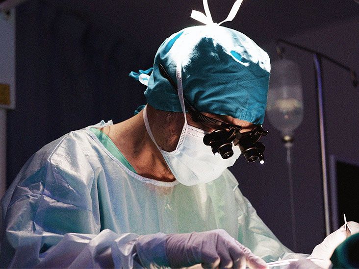
Retrosigmoid craniotomy sounds like a term invented by a sleep-deprived medical student with a Latin dictionary and too much coffee. In plain English, it is a skull-base brain surgery performed through an opening behind the ear, giving surgeons access to the area where the brainstem, cerebellum, hearing and balance nerves, facial nerve, and several important blood vessels share a very small neighborhood.
This approach is commonly used to treat conditions in the cerebellopontine angle, a deep space near the back of the brain. Doctors may recommend it for vestibular schwannoma, also called acoustic neuroma, certain meningiomas, microvascular decompression for trigeminal neuralgia or hemifacial spasm, and selected vascular or skull-base lesions. The goal is simple to say but technically demanding to do: reach the problem while protecting the nerves and brain structures that help you hear, balance, swallow, smile, blink, and feel your face.
If you or someone you love is preparing for this surgery, the medical vocabulary can feel like being handed a restaurant menu written entirely in neurosurgeon. This guide explains the purpose of retrosigmoid craniotomy, how the procedure is usually performed, what recovery may look like, and what questions to ask before surgery.
What is a retrosigmoid craniotomy?
A craniotomy is an operation in which a surgeon temporarily removes a small section of skull to reach the brain or nearby structures. “Retrosigmoid” describes the location: behind the sigmoid sinus, a large vein that drains blood from the brain. The incision is usually made behind the ear, and the bony opening is created in the lower back portion of the skull.
The retrosigmoid approach is also sometimes called the suboccipital approach. It allows the surgeon to work near the cerebellum and brainstem without going through the inner ear. That matters because, in some cases, this route may offer a chance to preserve hearing, especially when the hearing nerve is still functioning and the tumor or compression is suitable for this approach.
Why is retrosigmoid craniotomy done?
The purpose of a retrosigmoid craniotomy depends on the condition being treated. It may be used to remove a tumor, relieve pressure on a cranial nerve, treat a vascular abnormality, or obtain safe access to a deep skull-base area.
Vestibular schwannoma or acoustic neuroma
One of the most common reasons for a retrosigmoid craniotomy is removal of a vestibular schwannoma, often called an acoustic neuroma. This is usually a slow-growing, noncancerous tumor that develops on the balance and hearing nerve. Symptoms may include one-sided hearing loss, ringing in the ear, dizziness, balance problems, facial numbness, or, with larger tumors, pressure on the brainstem.
For acoustic neuroma surgery, the retrosigmoid approach can give the surgeon a broad view of the tumor, brainstem, facial nerve, hearing nerve, and surrounding blood vessels. In selected patients, it may allow hearing preservation attempts, although hearing outcomes depend on tumor size, tumor location, preoperative hearing level, nerve condition, and surgical factors.
Microvascular decompression
Retrosigmoid craniotomy is also used for microvascular decompression, often shortened to MVD. This procedure treats conditions caused by a blood vessel irritating a cranial nerve. Examples include trigeminal neuralgia, hemifacial spasm, and glossopharyngeal neuralgia.
During MVD, the surgeon looks for a blood vessel pressing against the nerve and places a tiny cushion between the vessel and nerve. The idea is not to remove the nerve but to stop the “pulsing poke” that can trigger lightning-like facial pain or involuntary facial spasms. Think of it as putting a polite little pillow between two neighbors who have been arguing for years.
Cerebellopontine angle tumors and other lesions
The same surgical corridor may be used for certain cerebellopontine angle meningiomas, epidermoid tumors, arachnoid cysts, cavernous malformations, and selected vascular conditions. The exact plan depends on imaging studies, symptoms, anatomy, and whether the goal is complete removal, partial removal, decompression, biopsy, or long-term control.
Who may be a candidate?
A person may be considered for retrosigmoid craniotomy when imaging shows a lesion that can be safely reached from behind the ear and when surgery offers meaningful benefit compared with observation, radiation, medication, or another surgical approach.
Doctors consider several factors before recommending the procedure, including:
- Type, size, and location of the tumor or lesion
- Symptoms such as hearing loss, facial pain, facial weakness, dizziness, or balance problems
- Whether the brainstem is compressed
- Current hearing level and whether hearing preservation is realistic
- Age, general health, anesthesia risk, and other medical conditions
- Previous surgery or radiation
- Patient goals, occupation, lifestyle, and tolerance for risk
Not every patient with an acoustic neuroma or cranial nerve condition needs surgery right away. Small, slow-growing tumors may be monitored with MRI scans. Some patients may be better candidates for stereotactic radiosurgery. Others may need a different surgical route, such as translabyrinthine or middle fossa craniotomy. Neurosurgery is not a “one size fits all” hoodie; it is tailored.
How to prepare before surgery
Preparation usually begins with a detailed review of MRI or CT scans, hearing tests, balance evaluation, neurological examination, and medical clearance. For acoustic neuroma, an audiogram is especially important because it helps determine whether hearing preservation is a reasonable goal.
Your care team may ask you to stop certain medicines before surgery, especially blood thinners, aspirin, or anti-inflammatory drugs, depending on your health history. Never stop a prescribed medication on your own; the surgeon and prescribing doctor should guide that decision.
Before surgery, ask about the expected length of hospital stay, incision care, pain control, activity limits, driving restrictions, return-to-work timing, follow-up imaging, and warning signs that require urgent help. Bring a written question list. The brain may be the star of the show, but paper still wins when nerves make your memory act like a browser with 47 tabs open.
What happens during a retrosigmoid craniotomy?
The operation is performed under general anesthesia. After you are asleep, the surgical team positions your head carefully to give the surgeon a safe angle to the area behind the ear. Hair may be clipped around the incision site. The exact amount varies by hospital and surgeon; modern teams often avoid shaving more than necessary.
The surgeon makes an incision behind the ear and creates a small opening in the skull. The protective covering of the brain, called the dura, is opened. The cerebellum is gently relaxed or retracted just enough to create a working corridor. Surgeons use a microscope, microsurgical instruments, nerve monitoring, and image guidance when appropriate.
If the surgery is for a vestibular schwannoma, the tumor is carefully separated from surrounding nerves and blood vessels. Sometimes a small piece of tumor may be intentionally left behind if removing it would risk serious nerve injury. In that situation, the remaining tissue can be monitored or treated later.
If the surgery is for microvascular decompression, the surgeon identifies the affected nerve, finds the blood vessel causing compression, and places a small pad between them. After the main work is complete, the dura is closed, the bone opening is repaired or covered, and the incision is closed with sutures or staples.
How long does surgery take?
The length of a retrosigmoid craniotomy varies widely. Microvascular decompression may take several hours. Tumor surgery may take longer, especially if the tumor is large, firm, stuck to nerves, or close to the brainstem. Time in the operating room also includes anesthesia, positioning, monitoring setup, and closing.
A longer surgery does not automatically mean something went wrong. Skull-base surgery is delicate work. Nobody wants a neurosurgeon rushing like they are trying to beat airport boarding.
What to expect after surgery in the hospital
After surgery, many patients spend time in a recovery unit or neuro-intensive care setting for close monitoring. Nurses and doctors check alertness, strength, eye movement, facial movement, hearing changes, balance, swallowing, pain, nausea, and the incision site. You may have an IV line, monitors, compression devices on your legs, and medication for pain or nausea.
Headache, neck stiffness, fatigue, nausea, dizziness, and swelling around the incision are common after a craniotomy. Some people feel surprisingly decent; others feel as if their head has filed a formal complaint. Both experiences can be normal, but symptoms should gradually improve.
Hospital stays vary. Some patients go home after a few days, while others stay longer depending on the reason for surgery, symptoms, balance, pain control, complications, and support at home.
Recovery timeline after retrosigmoid craniotomy
First few days
The early focus is safety: controlling pain, preventing nausea, walking with help, checking neurological function, and making sure the incision is healing. Balance may feel off, especially after vestibular schwannoma surgery. Physical therapy or vestibular therapy may begin in the hospital if needed.
First two weeks
At home, patients usually need rest, short walks, incision care, and help with meals, transportation, and chores. Heavy lifting, bending, straining, and intense activity are usually restricted. Stitches or staples may be removed during a follow-up visit, depending on the surgeon’s instructions.
Weeks three to eight
Energy often returns slowly. Many people can increase walking and light daily activities, but fatigue may arrive suddenly, like a phone battery going from 38% to “absolutely not.” Some patients return to desk work in several weeks; others need more time, especially after larger tumor surgery or if dizziness, facial weakness, headaches, or balance issues persist.
Longer-term recovery
Full healing can take months. Nerves recover slowly, and some symptoms may improve gradually over time. Follow-up may include MRI scans, hearing tests, facial nerve evaluation, balance therapy, or additional treatment if a tumor remnant is being monitored.
Possible risks and complications
Every brain surgery has risks, and the retrosigmoid approach is no exception. Your surgeon should explain risks based on your diagnosis and anatomy.
Possible complications include:
- Bleeding, infection, or reaction to anesthesia
- Cerebrospinal fluid leak
- Headache or persistent incision-area pain
- Hearing loss or tinnitus
- Dizziness, vertigo, or balance problems
- Facial weakness or facial paralysis
- Facial numbness or changes in taste
- Swallowing or voice changes, depending on nerve involvement
- Stroke, blood clots, seizures, or brain swelling, though these are less common
- Need for rehabilitation or additional treatment
Risk is influenced by tumor size, tumor attachment to nerves, prior treatment, medical history, surgeon experience, and the specific condition being treated. For acoustic neuroma, facial nerve preservation is often a major priority. For microvascular decompression, the goal is symptom relief while avoiding nerve injury.
When to call the doctor after surgery
Call your care team promptly if you develop fever, worsening headache, increasing redness or drainage from the incision, clear fluid leaking from the wound or nose, new weakness, confusion, seizure, severe vomiting, worsening balance, shortness of breath, chest pain, or new facial drooping. If symptoms feel sudden or life-threatening, seek emergency care.
It is better to make one cautious phone call than to silently negotiate with a symptom that clearly did not come to the table in good faith.
Questions to ask your surgeon
Before retrosigmoid craniotomy, consider asking:
- Why is the retrosigmoid approach best for my condition?
- Are observation, radiation, medication, or another surgical approach reasonable options?
- What are the goals: complete removal, partial removal, decompression, or symptom relief?
- What are my chances of hearing preservation or facial nerve preservation?
- How many similar procedures does the team perform?
- How long might I stay in the hospital?
- What symptoms are expected, and which symptoms are urgent?
- Will I need vestibular therapy, facial therapy, hearing support, or follow-up MRI scans?
- When can I drive, work, exercise, fly, or lift heavy objects?
Practical recovery tips
Plan your recovery space before surgery. Put frequently used items at waist height so you do not need to bend. Prepare soft, easy meals. Arrange rides to appointments. Ask one trusted person to help track medications, discharge instructions, and follow-up dates.
Walking is often encouraged, but follow your surgeon’s limits. Short, frequent walks are usually better than one heroic expedition to prove you are “fine.” Healing is not a reality show. There is no trophy for overdoing it.
Protect your sleep. Brain recovery loves rest. Keep hydration steady, avoid constipation, and take pain medicine exactly as directed. If dizziness is present, use handrails, remove trip hazards, and ask about vestibular rehabilitation.
Experiences related to retrosigmoid craniotomy: what patients often notice
Many people preparing for a retrosigmoid craniotomy want to know what the experience feels like beyond the textbook description. While every patient is different, several themes come up again and again in recovery conversations.
First, the emotional build-up before surgery is often harder than people expect. A person may understand the medical reason for surgery and still feel scared, restless, or strangely calm one hour and panicked the next. That is normal. Brain surgery is not the same as getting a cavity filled. Patients often find it helpful to ask the surgeon to explain the plan in plain language: where the incision will be, what structures are being protected, what outcome is realistic, and what would make the team change plans during surgery.
Second, the first days after surgery can feel disorienting. Patients may wake up with a sore neck, a tight bandage, a dry mouth, and more wires attached than a home entertainment system from 2007. Nurses may ask the same neurological questions repeatedly: name, date, location, smile, squeeze hands, follow a finger. It can be annoying, but those checks are important. They help the team spot changes early.
Third, balance can be a surprise. After retrosigmoid surgery for vestibular schwannoma, some patients feel wobbly when they first stand or turn their head. The brain usually adapts, but it may need practice. Vestibular therapy can teach safe movements and exercises that help the brain recalibrate. Progress may be slow at first, then suddenly better, then temporarily worse after a busy day. Recovery does not always move in a straight line; sometimes it prefers interpretive dance.
Fourth, fatigue is real. People may look “normal” but feel exhausted after a shower, a short walk, or a conversation with relatives who mean well but ask fifteen questions in one breath. Rest periods are not laziness. They are part of neurological healing. A good rule is to increase activity gradually and stop before the body stages a dramatic protest.
Fifth, incision sensations can be odd. Numbness, itching, tightness, or brief zaps near the incision may occur as tissues heal. Patients should still report worsening pain, swelling, fever, drainage, or fluid leakage. Normal healing can feel weird; infection or cerebrospinal fluid leak needs medical attention.
Finally, recovery often includes a “new normal” phase. Some people return quickly to work and daily routines. Others need hearing support, balance therapy, facial exercises, or more time to regain confidence. The best experience usually comes from clear expectations, a skilled multidisciplinary team, honest communication, and patience. Retrosigmoid craniotomy is a major procedure, but for the right condition and the right patient, it can relieve pressure, remove disease, protect vital nerves, and improve quality of life.
Conclusion
Retrosigmoid craniotomy is a specialized skull-base operation that gives surgeons access to the area behind the ear near the cerebellum, brainstem, and cranial nerves. It is commonly used for vestibular schwannoma, microvascular decompression, cerebellopontine angle tumors, and selected vascular lesions. The procedure can be highly effective, but it requires careful planning because the surgical target sits close to structures that control hearing, balance, facial movement, facial sensation, swallowing, and coordination.
Patients should understand why this approach is being recommended, what alternatives exist, what risks apply to their specific case, and what recovery may involve. With realistic expectations, good follow-up care, and a recovery plan that respects the brain’s need for rest, many people move through surgery and healing with confidence rather than confusion.
Note: This article is for educational purposes only and does not replace medical advice. Diagnosis, treatment choice, surgical risk, and recovery expectations should always be discussed with a qualified neurosurgeon or skull-base specialist.